Monday, January 12, 2009

POXVIRIDAE

Structure of the Poxviridae:


Genome
- Large double stranded DNA about 130-300 kbp in size.
- Linear genome.
- Noninfectious
- The ends of the genome form direct repeats known as inverted terminal repeats (ITRs).
- The important genes can be found in the central part of the genome whereas the non-essential genes are located at the ends of the genome.

Large DNA viruses
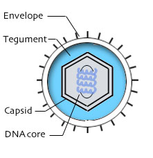
- About 200-400 nm long.
- The particles are very complex and covered with filamentous protein components that contain more than 100 proteins.
- Two morphological types of particles:
(i) Oval with criss-cross surface bonding
(ii) Brick-shaped
d. The whole particle is enclosed with envelope from host cell membrane.

Poxviruses have a complex structure.
- It can induce the specific and cross reacting antibodies.
- That is why it has the ability to vaccinate against one disease with other virus.

  • Poxviridae has two sub families: Chordopoxvirinae and Entomoxvirinae.
  • Chordopoxvirinae: Poxviruses of vertebrates.
  • Entomoxvirinae: Poxviruses of invertebrates more specifically insects.


  • Poxviruses that can infect humans can be found under the chordopoxvirinae sub family and most viruses can be found from the orthopoxvirus and the parapoxvirus genus.
  • Poxviruses are usually transmitted through direct contact.
  • Other poxviruses:
1. Vaccinia

Vaccinia belongs to the Orthopoxvirus genus is used as a vaccine against the smallpox virus. Since the vaccinia vaccine virus does not contain the smallpox virus, it cannot cause any smallpox infection. This virus cannot be transmitted through air (hence you cannot get infected by being in the same room as the infected person) but a person can be infected if the person has direct contact with the vaccination site. Some of the symptoms include rash, fever, headache and body ache.


From left:
http://upload.wikimedia.org/wikipedia/commons/8/8c/Vaccinia_virus_PHIL_2143_lores.jpg
http://www.bt.cdc.gov/training/Smallpoxvaccine/reactions/gen_vac_clinical.html


2. Smallpox virus

Smallpox, a member of the Orthopoxvirus is a severe, contagious disease that is caused by the variola virus. This virus believed to be originated in India or Egypt and it was the most fatal disease during 3000 years ago as there was no cure for the virus even though a major population of the people was infected with it. Smallpox can be spread through direct contact with contaminated things like clothing. The initial symptoms are high fever, headaches and body aches before progressing to rash.


3. Orf virus

It is a virus that belongs to the parapoxvirus genus. This orf virus causes infection to sheep and goats and sometimes the virus can infect to people that handle the sheep and goats. This is because the virus can establish infection where the skin is damaged. Painful lesions were seen first before it increases in size over a period of 4 weeks. Then the lesion will crust before it dries up after 6 to 8 weeks and fall off leaving with no scars.


From left:
http://www.cdc.gov/ncidod/dvrd/orf_virus/images/orf_kid_lg.jpg
http://upload.wikimedia.org/wikipedia/commons/a/a2/Orf_virus_infection_on_thumb.jpg


References:

Sunday, January 11, 2009

ORTHOMYXOVIRIDAE

Here is a mindmap of ORTHOMYXOVIRIDAE based on the lecture notes.


Wednesday, January 7, 2009

PICORNAVIRIDAE

Here is a mindmap on Picornaviridae based on the lecture notes:


Tuesday, January 6, 2009

HERPESVIRIDAE

There are 8 types of Herpes virus:


  • Herpes viruses quite ubiquitous in nature (ubiquitous means everywhere) and they are prevalent (means common/widespread).
  • Herpes viruses are the leading cause of human viral disease just second to the influenza virus.
  • Herpes viruses can be found mammalian and non mammalian vertebrates and non vertebrates.
  • Structure of Herpes virus:


from left:

http://stdgen.northwestern.edu/stdgen/bacteria/hhv2/herpes.diagram.jpg

http://stdgen.northwestern.edu/stdgen/bacteria/hhv2/herpes.diagram.jpg

- Envelope: Found in the outside of the herpes virus particle. Contain numerous glycoproteins.

- Tegument: It is the space between envelope and the capsid. The tegument is a protein-filled region which contains protein and enzyme which are involved in the beginning of the replication process.

- Glycoprotein spikes: It determines the cell that can be infected due to the availability of the suitable receptors.

- Capsid: Protein shell of the herpes virus. It has an icosahedral structure.

- Genome: Contains linear double stranded DNA.

Herpes Simplex Virus 1 (HSV1) and Herpes Simplex Virus 2 (HSV2):

Similarities
- Both share about 50% of their DNA.
- Most people infected with HSV1 or HSV2 do not have any symptoms or any noticeable symptoms.
- Both infect the mucosal surfaces of the body.

Differences
- HSV1 causes the sores around the mouth and it is also known as “cold sores”.
- HSV2 infects the genital and it is also known as “genital herpes”.
- HSV1 establish latency in the trigeminal ganglion.
- HSV2 establish latency in the sacral ganglion.

Causes of HSV1 and HSV2
- People get infected with HSV1 by touching infected saliva mucous membrane or skins.
- For HSV2, people get infected during sex with an infected partner.

Stages of Infection

  1. Primary infection
    When people get infected with either HSV1 or HSV2, they usually do not know of the condition since most of the time there are no symptom or noticeable symptom.
  2. Latent infection
    After the herpes viruses infect the host, it will remain inactive for a long period of time. It hides within in the cells and the nerves tissue. By hiding, the herpes virus protects itself from being destroyed by the antibodies that is produced by the immune system.
  3. Recurrent infection
    This happens when the herpes virus reactivates. Reactivation occurs due to various factors like sunlight, stress and menstruation. Infections can recur throughout life.

References:

HEPADNAVIRIDAE

  • Hepadnaviridae is obtained from the hepatotrophic (replicates and cause infection of the liver) nature of the virus and its genome which is made up of double stranded DNA (hepadna).
  • All hepadna viruses can cause hepatitis to their host as they infect hepatocytes that can be found in the liver.
  • Hepatitis: Syndrome characterized by inflammation of the liver.
  • Although all hepadnaviruses cause hepatitis to their host, but this does not mean that all hepatitis diseases are hepadna.
  • Other hepatitis include:
    - Hepatitis A (Picornaviridae)
    - Hepatitis C (Flaviviridae)
    - Hepatitis D (No family)
    - Hepatitis E (Hepeviridae)
    - Hepatitis G (Flaviviridae)


from left in the top row:


Hepatitis A (http://www.marlerblog.com/gech_0001_0002_0_img0129(1).jpg)
Hepatitis C (http://www.co.monroe.mi.us/Monroe/uploadedImages/hepatitis-c(1).gif)
Hepatitis D (http://www.dshs.state.tx.us/idcu/disease/hepatitis/hepatitis_d/image1.gif)
Hepatitis E (http://pathmicro.med.sc.edu/virol/hep-e1.jpg)
Hepatitis G (http://www.dshs.state.tx.us/idcu/disease/hepatitis/hepatitis_g/image1.gif)

  • Hepadnaviridae can be divided into two genera: Orthohepadnavirus and Avihepadnavirus.
  • The genera are divided based on the similarities of the DNA sequence.
  • The orthohepadnavirus infect mammals whereas the avihepadnavirus infect the avians meaning the birds.
  • These viruses infect species that are closely related to their natural host. Example hepatitis B viruses infect humans and chimps which are closely related. That is why they have a narrow range of hosts.


  • Among the virus classified under hepadnaviridae, only one virus that is pathogenic to human: Hepatitis B.

  • Structure of hepadna virus:
Genome:
a. Hepatitis B has the smallest genome of 3.2 kb long among all hepadna viruses.
b. Partially double stranded DNA genome which carries all the genetic information in it.
c. The DNA consists of a positive strand and negative strand.
d. The complete negative strand is longer than of the incomplete positive strand (about 15% to 50% of the DNA is single stranded).
e. An RNA oligonucleotide is attached to the 5’ end of the positive strand while a 5’ protein covalently attached to the end of the negative strand.

Nucleocapsid:
a. Has an icosahedral shape.
b. The nucleocapsid is about 27 nm in diameter.
c. Contains a DNA polymerase and an antigen (HBcAg)
d. Three proteins can be found on the surface of the capsid: L (large), M (medium) and S (small) that is present in the ratio of 1:1:4.


Envelope:
a. It is spherical and 42 nm in diameter.
b. It surrounded the nucleocapsid.
c. Contain glycoproteins, lipids and surface antigens that is virus-specific like HBsAg.


References:

Sunday, January 4, 2009

Classification of Viruses

viruses do not follow the taxonomy of bactera, fungi and animals..

Saturday, January 3, 2009

Overview of Virology

A Peek into VIROLOGY

There are 3 main domains of life:
  • Eukarya
  • Eubacteria
  • Archaea

However, viruses do not fall in any of the domains as they are not considered organisms however, they have life-like properties. They depend on other organisms for reproduction and metabolism.

Their common physical characteristics/properties are as follows.

  1. Intercellular parasites
  2. one type of nucleic acid (either DNA or RNA) - genome
  3. no metabolic activity outside host cells

Each virus particle has the following structures:

  1. virion - the intact virus particle
  2. capsid - protein coat (which protects the viral genome form inactivation by adverse environmental factors)
  3. capsomeres - capsid is composed to these (protein sttuctural units)
  4. nucleic acid
  5. envelope (many virus particles are surrounded by a lipoprotein envelope that contains viral antigens, partly derived from the outer membrane or in some cases, the nuclear membrane of the host cell)

There are 3 types of symmetry shown by virus particles:

  1. cubic - particles are icosahedral protein shells with the nucleic acid contained inside
  2. helical - particle contains an elongated nucleocapsid & the capsomeres are arranged round the spiral ofthe nucleic acid
  3. complex - particles do not conform to none of the above symmetry mentioned